- Home
- About ANT
-
Products
asa
asa is a highly flexible EEG/ERP and MEG analysis package with a variety of source reconstruction, signal analysis and MRI processing features.
.jpg)
eego mylab
The new frontier in multimodal brain research. With up to 16 kHz sampling rate, 256 EEG channels and unique software features, eego mylab gives you an unprecedented in-depth understanding of the human brain.
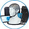
eego sports
eego sports offers complete freedom to collect high-density EEG data, bipolar EMG signals, and a variety of physiological sensor data, wherever and whenever required, with publish quality data in less than 15 minutes!
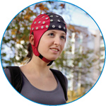
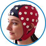
waveguard net
The waveguard net sets a new standard for research applications requiring high-density EEG data acquisition with quick preparation time, high flexibility, and subject comfort.
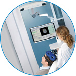
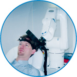
visor2
Our new and upgraded visor2 solutions integrate all the latest technologies for navigated rTMS, dual-coil navigation support, EEG-TMS recordings and pre-surgical evaluation for the highest quality in research and clinical procedures.
powerMAG ANT
The PowerMAG ANT 100 rTMS stimulator is designed for the specific needs of high-end TMS applications. Powerful high-frequency TMS as well as high precise single pulse and repetitive pulse protocols are combined in one single device.
xensor
xensor offers the solution for digitization of 3D electrode positions. xensor takes care of the whole procedure; it records, visualizes and stores positions acquired with a dedicated digitizer.
waveguard original
waveguard original is the cap solution for EEG measurements compatible with fMRI, MEG and TMS system. Use of active shielding guarantees performance in even the most demanding environments.
waveguard connect
waveguard connect EEG caps are a perfect match for hospitals and institutes aiming at reliable EEG, maximum uptime and great patient comfort! For optimal signal quality, the electrodes are made of pure, solid tin.
waveguard touch
waveguard touch is a dry electrode EEG cap. The unique Ag/AgCl coated soft polymer electrodes provide stable, research-grade EEG signals while maintaining subject comfort. The combination of these innovative dry electrodes and the industry-leading waveguard cap makes waveguard touch the best solution for dry EEG.
smartmove
smartmove allows planning of a complete TMS session ahead by defining stimulation sites based on anatomical MRI information and functional information like fMRI, PET or EEG/MEG.
- References
- Support
- Events
- News
- Contact Us
 Read more
.jpg)

You are here
MEG source analysis with asa
MEG source analysis with asa
asa was showcased in an interactive workshop by Dr. Susan Bowyer, Ph.D. at the American Clinical Magnetoencephalography Society (ACMEGS) annual meeting. The meeting took place in San Antonio, Texas from February 6-12, 2012. Dr. Bowyer presented MEG analysis using asa, on cases with Evoked Eloquent Cortex Responses.
The data was recorded with a whole head 4D Neuroimaging (BTi) MEG system. After importing the data into asa, the data were band-pass filtered between 3 and 50 Hz. Next, the data were averaged and finally, a latency marker was placed at the peak that would be used for source analysis.
As a preparatory step for source analysis, a 3D (BEM) head model was created with segmentation using the patient`s individual MRI.
Dr. Bowyer used a combination of swLORETA and dipole fit for her presentation on MEG source analysis in asa. For Dr. Bowyer, the swLORETA calculated an initial solution, visualized in data-output and in the 3D brain-space. She then automatically fed the maximum of this solution into the dipole fit algorithm. The swLORETA was calculated first in order to prevent the dipole fit algorithm from getting stuck at a local maximum. The a priori information coming from the maximum result of the swLORETA solution not only avoids the unlikely but undesirable pitfall, it also ensures that the dipole fit outputs the highest possible localization accuracy.
The presented showcase contained data of a 15 years old female who had history of intractable right arm simple parietal sensory seizures, sometimes accompanied by contractions of the right arm muscle from elbow to wrist. She had a left parietal tumor removed 5 months prior to MEG study. She continued to have sensory seizures in her right arm from 0-5 times per day. MEG was performed to plan a second surgery, such as multiple subpial transections into the epileptogenic focus to relieve these sensory type seizures. The MEG and EEG recordings were abnormal and showed focal slowing seen in the left parietal and temporal regions.
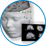
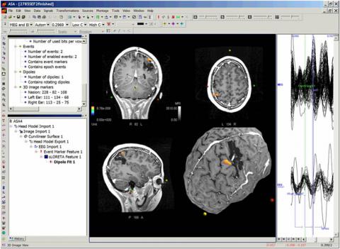
The resulting image below is from the final output of Dr. Bowyer`s data and was used in her presentation. Averaged traces are shown on the rightmost window. In the center window, one can see the dipole fit result visualized in the MRI slices as well as on the curvilinear surface. The dipole was calculated on somatosensory evoked fields (SEF) and localized to the post central gyrus in the anterior margin on the tumor resection.
About Dr. Susan Bowyer
Dr. Susan Bowyer, Ph.D. is a medical physicist and neuroimaging scientist in the Neuromagnetism Lab of Henry Ford Hospital in Detroit, Michigan.
About asa
The software package integrates all current MEG file formats and allows users to open and read MEG/EEG data without the need to export the files. asa supports a wide range of data formats for import and analysis, including the 4D BTi format, and other the CTF and Neuromag MEG data formats. Additionally, the software automatically reads the 3D positions of the SQUID (Superconducting Quantum Interference Device) magnetometers. Apart from creating individual head models, the asa software comes with the MNI (Montreal Neurological Institute)-standard and Collins-standard brains.
MUSIC metric to estimate regions with high likehood of focal electrical activity.


ANT Neuro
Welbergweg 74
7556 PE Hengelo
Netherlands
T: +31 (0) 85 049 8175
F: +31 (0) 85 049 3919
E: Send us an email